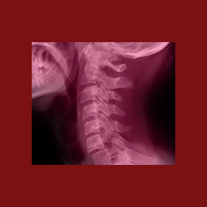
Facet joint x-ray testing can provide a detailed look at the skeletal structures of the apophyseal joints. While x-ray is helpful towards the diagnosis of some facet joint issues, it is limited in effectiveness due to the simple fact that it can not visualize soft tissues, inflammation or other common causes of facet syndrome. Therefore, although x-rays have their uses, they are not ideal as an enlightened facet syndrome diagnostic tool.
X-ray technology is the oldest form of medical imaging and has been used since the late 1800s to depict the inner workings of the human skeletal structure. Many facet joint problems are caused by osteophytes, which are simply outcroppings or accumulations of bony material. Therefore, these bone spurs will image well on x-ray films and might provide insight as to the reason why a given patient might experience zygapophyseal joint pain. However, as this website explains, there are many possible causes of facet joint pain and some of these contributing factors do not involve the skeletal system. Therefore, x-rays are limited in their application for diagnosing facet joint pathologies and should be used only when access to better methods is absent or restricted.
This critical essay examines the modern use of x-ray technology to diagnose facet syndrome, including the use of live fluoroscopy during diagnostic injection and surgical treatment procedures.
Facet Joint X-Ray Imaging
X-ray images have been used for more than a hundred years to visualize the interior skeletal components of the body. Since bone matter is dense, it contrasts against skin and other soft tissues, providing very vivid images. X-rays can be focused to inspect a small region of the spine, such as specific facet joints and can prove the existence of bone spurs, ligamentous ossification and foraminal narrowing caused by facet arthritis and degeneration.
General purpose x-rays typically are used for preliminary evaluation of back and neck pain complaints. This means that they will image larger areas of the anatomy in less detail. Once specific regions can be concentrated upon, the images can provide greater detail of smaller anatomical targets. However, the resolution of x-ray can not compare to the quality of images produced by more advanced technologies, such as CT scan and especially MRI.
X-Ray Fluoroscopy
Fluoroscopy is a form of live x-ray that does not capture images on film, but instead displays them on a monitor so that the doctor can observe the interior of the skeletal system in real time. Fluoroscopy has many uses and we will detail the ones that are specific to facet syndrome treatment below:
Fluoroscopy can be used to observe the painful facet joint as it moves. This might provide insight as to the mechanical nature of pain by observing osteophytes that may interfere with the range of motion in the affected joint.
Fluoroscopy is a commonly used guidance tool for diagnostic injections. The theory of suspected facet joint pain is often confirmed by using an anesthetic injection into the joint. If the pain diminishes or resolves, then that particular joint can be “confirmed” as the origin of symptomatic activity. Fluoroscopy improves the accuracy of these diagnostic injections and should always be utilized when this form of facet joint evaluation is performed.
Fluoroscopy can also be very helpful during surgical techniques. Live imaging allows the surgeon to visualize much of the greater spinal anatomy without having to utilize large incisions or tissue dissection to observe the structures directly. Fluoroscopy can improve the results of many surgical techniques and works well in combination with other visualization practices such as fiber optics, laser optics and various catheter-based methods of care.
Facet Joint X-Ray Benefits and Limitations
X-rays can accurately and comprehensively detail skeletal changes in the facet joints. They can visualize bone spurring, hypertrophy of the skeletal joints, ligament ossification and arthritic neuroforaminal stenosis. These are all helpful additions to any diagnostic evaluation. X-rays are inexpensive and generally available from doctors, chiropractors and other types of care providers.
On the downside, x-rays are limited in what they can image. They are not helpful in identifying soft tissue joint inflammation in or around the facet joints or most forms of ligament-related joint dysfunction. Additionally, x-rays demonstrate some degree of health risk, especially from repeated exposure in the same area of the body.
We find the best use of x-ray technology to be that of fluoroscopy. This live form of imaging is very helpful for improving the accuracy, efficacy and safety of injection and surgical techniques and should be implemented during any of these medical endeavors in order to provide the best outcomes.
Facet Joint Pain > Facet Syndrome Diagnosis > Facet Joint X-Ray


