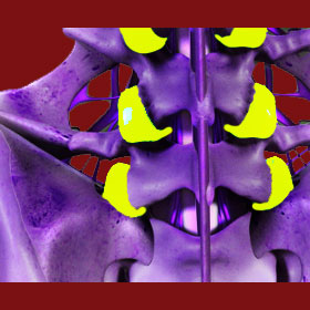
Facet joints are important parts of the dorsal anatomy, helping to connect the vertebrae together and stabilize the overall length of the spinal column. Other common medical terminologies for these vertebral joints include the names zygapophyseal joints or apophyseal joints. However, these nomenclatures are rarely used outside of research science and will virtually never be utilized by diagnosticians when communicating with patients due to their complexity of pronunciation, as well as multiple common spellings. “Facet” is most popular name for these spinal connectors.
Facet joints have become a common topic of discussion between various types of care providers and their patients. Degeneration, hypertrophy and injury to these structures is theorized to be a major source of back and neck pain, also known medically as dorsalgia or dorsopathy. Although well known to students of anatomy for thousands of years, the zygapophyseal joints have now become mainstream as more and more patients are diagnosed with various forms of pathology governing facet form and function. The diversity of possible pain conditions that have been linked to the spinal facet connectors is known as facet syndrome.
This background resource section covers the anatomy of the spinal apophyseal joints. We will examine the way that the facets are designed, how they operate and what purposes they serve in the human vertebral column.
Anatomy of the Facet Joints
Each facet is comprised of a meeting pair of articular processes from neighboring vertebrae. There are one set of facet joints connecting each vertebral level of the backbone. Each set contains 2 facets placed bilaterally on the left and right sides of the spinous process, which is the large central projection that is palpable on many of the vertebrae of the spine. The facet joints form at the juncture of the superior and inferior articular processes. These are the small upward and downward facing projections flanking the spinous process. The superior process is found on the top side of most vertebral bones, while the inferior process is located on the bottom side of most vertebral bones.
Each facet is covered in cartilage and is surrounded by a capsule of protective fluid, encased by ligaments. All together, this design is called a synovial joint and is found in many anatomical locations in the human body. The cartilage protects the bony articular projections of the joint, while the synovial fluid helps to lubricate the joint against friction and interaction between bony components. The ligaments hold the joint capsule together and regulate the tension and laxity of the joint in regards to movement tolerances.
Apophyseal joints connect the superior and inferior articular processes of 2 vertebrae from one spinal level to the next. This forms a continuous network of joinings that are referred to as the articular pillars of the backbone. The 4 main spinal ligaments all work to provide structural support for the facet joints, including the posterior longitudinal ligament, the ligamentum flavum, the interspinous ligament and the superspinous ligament.
Spinal Joint Topics
Below are all of our topical discussions on the subject of apophyseal joints. As new essays are published they will be added to the section below:
Facet joint osteophytes and also commonly known as bone spurs. These small skeletal projections are the results of the osteoarthritic processes and are normal to exist throughout many areas of the back bone.
Facet hypertrophy describes an inflammation of the zygapophyseal joint structure, creating an abnormally large appearance.
Facet degeneration is a universal result of spinal aging. Degeneration is not inherently pathological and should never be assumed to be the source of any symptoms unless pathology can be firmly established.
Are you still seeking more information? Learn even more in our detailed analysis answering the question: What are facet joints?
Function of the Facet Joints
The spinal zygapophyseal joints serve several important functions and can produce pain and other symptoms when their functionality decreases due to injury, pathological deterioration or dislocation:
The facets literally connect each vertebral bone to the one above and below. This physical bond is one of the main sources of stability in the vertebral column and allows the entirety of the backbone to be far stronger than each component alone.
The apophyseal joints help to regulate movement in each individual vertebral level and collectively, in the entire spinal column. The joints help to provide safe range of motion for flexion (bending forward or anteriorly) and extension (bending backwards or posteriorly), as well as inhibit excessive vertebral rotation. Rotational limitation is one of the most specific functions of the zygapophyseal joints.
The facet structures also help to maintain and regulate spinal alignment and positioning of each vertebral bone in relation to the others which surround it.
Facet Joint Pain > Facet Joints


